The termite mound: A not-quite-true popular bioinspiration story

The termite mound: A not-quite-true popular bioinspiration story

I traveled extensively over the summer; to Austin, TX, all through Western Europe and back to Illinois. All the while I was working on this blog post about bioinspired air-conditioning, which was appropriate because everywhere I went I seemed to have to suffer through heat-wave after heat-wave.

While wishing for Europe to have more air-conditioning units (especially in class-rooms and lecture halls), one of course wonders if that would only exacerbate the problem and make summers even hotter. Progress is constantly being made on making air-condition units more compact, more energy efficient, and thus more environmentally friendly. Inspiration on how to accomplish this has been already been found in natural systems.

Recently Brian Clark Howard wrote an interesting and popular article for National Geographic entitled: “5 Natural Air-Conditioning Designs Inspired by Nature”. Arthropods (termites & ticks) were prominently represented on this list. However, I would like to provide a little bit more detail and corrections to the NG’s list.

The insect examples touched upon in the NG article is that of the termite mound. The most famous architectural example of biomimicry or bioinspiration is the Eastgate Centre Building in Harare, Zimbabwe, which opened in 1996. Architect Mick Pearce and the firm Arup were supposedly partly inspired to build a building suitable for a tropical climate by considering the locally present termite mounds, and build their vision using locally available materials.

Eastgate Centre (with chimneys on roof) in Harare Zimbabwe. (Source: wikipedia.org. Unknown potographer)

Eastgate Centre (with chimneys on roof) in Harare Zimbabwe. (Source: wikipedia.org. Unknown photographer)

Anyone who is even the slightest bit interested in biomimicry knows of the Eastgate Centre, but since the political and economic situation in Zimbabwe has probably only deteriorated since 1996 I was wondering if it was actually still standing and still used as the commercial center it had been envisioned as.  So I took my questions to Twitter: [View the story “EastGate Building Harare, Zimbabwe” on Storify]. Turns out that the Eastgate building is still used and stands out prominently in the heart of Harare (and as @ardeans pointed out the building is located right on Robert Mugabe Road, sigh). One of the occupants is actually the United States Embassy.

It is just kind of too bad that the building is based on incorrect biology. Or is it?

First a little bit of background about the inspirational insect. Macrotermes termites (Macrotermitinae) occur over tropical Africa and Asia. There are about 330 species in this genus of relatively large termites. Most of the species build elaborate mounds. The tallest mounds occur in Africa (max of 30 feet, 9 meters). Macrotermes termites cultivate fungi, and spend most of their time, somewhere deep within the mound. The Macrotermes species that has been studied the most is the African species M. michaelseni.

Termite Mound

Flickr user Potjie uploaded this picture of a termite mound in Northern Namibia. (http://www.flickr.com/photos/potjie/3408013097/)

Popular wisdom says that in order to optimize fungus growth the interior of the mound needs to be maintained within the narrow range of 29-32°C. To keep keep within this narrow temperature range, despite of fluctuating temperatures during the day- and night time, the large mounds are not just heaps of dirt but in fact incorporate elaborate ventilation holes and air ducts and air pockets, which drive natural ventilation through convection. And remember, these structures are build by a million tiny insects that behave in an organized manner to come up with an architectural masterpiece, every time.

So no wonder that Mick Pearce, born and raised in Southern Africa, was inspired by the the termite mound to design a building in tropical Harare that would have similar features.

Up until recently two models for the termite mound function were proposed and commonly accepted. In case of mounds that were capped a “thermosiphon flow” was created – basically hot air created by the nest rises to the top of the mound where it gets refreshed and is supplied with water vapor through the porous mound walls. This denser air then is forced down below the nest, where the cycle is repeated.

2013-09-04 08.40.10

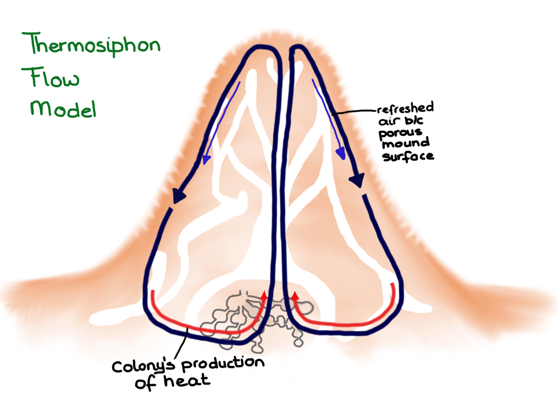
Thermosiphon flow model in which ventilation is driven by heat. (Drawing by Marianne Alleyne, based on Turner & Soar, 2008)

The second model applies to mounds that have a chimney at the top of the mound that opens to the outside. This arrangement creates induced flow, also called the stack effect. The chimney breaks the surface boundary layer and is exposed to higher wind speeds compared to inlets on the ground. The unidirectional flow draws fresh air from near the ground into the nest, where it passes on through the chimney and ultimately to the outside.

2013-09-04 08.40.20

Induced flow model in which ventilation is driven by wind. (Drawing by Marianne Alleyne, based on Turner & Soar, 2008)

The architects who designed the Eastgate Centre building tried to incorporate both the thermosiphon and induced flow principle into their design. The building has an extensive tube system within the walls and floors that move air trough the building. Heat generated within the building, along with stored heat within the structure, creates a thermosiphon-effect that draws air up and through the rooftops where large chimney stacks are located. These tall stacks are essential for creating an induced flow.

Detail of tube system within Eastgate Centre building.

Detail of tube system within Eastgate Centre building’s walls and floors (Original picture at http://harare.usembassy.gov).

When popular stories about the Eastgate Centre building say that the building works on the termite mound principle they ignore the fact that the building uses low capacity fans during the day, and high capacity fans during the night to keep the air from being too stagnant, effectively replacing the hot air that builds up during the day with cool air during the night. This works well, and avoids having to use expensive air-conditioning technology, but needless to say, no termite mound utilizes fans.

DayNight

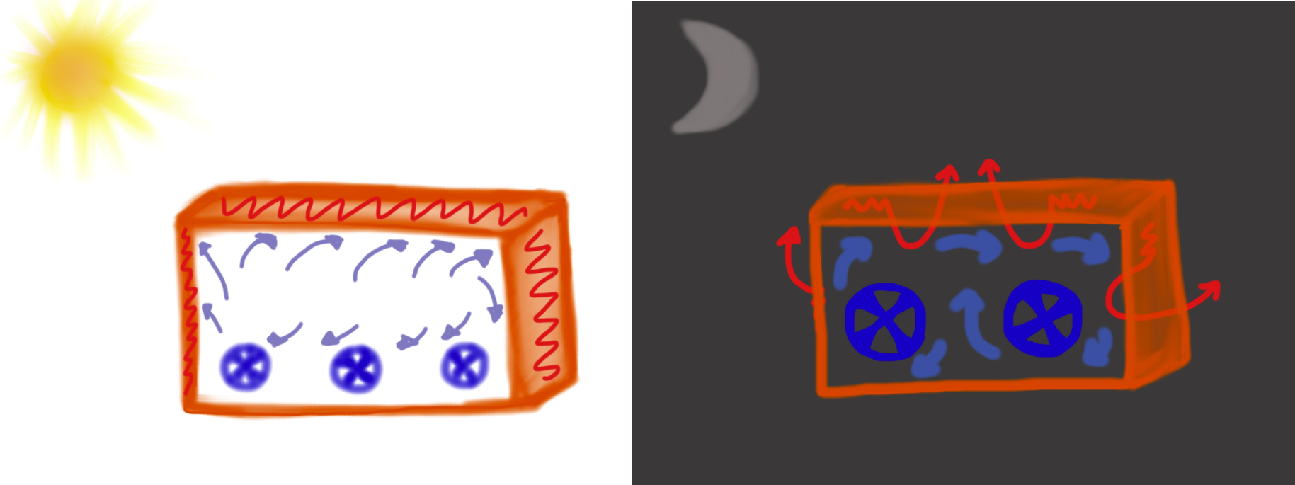
The use of fans within the Eastgate Centre. Smaller fans run during the day-time hours (left). They keep the environment within the building comfortable while the walls store the heat from the outside. Larger fans run during the night-time (right), these fans pull the stored heat out of the walls and push the heat out through the ducting in the ceiling and walls. By the next morning the walls are ready to again store heat (Drawing by Marianne Alleyne).

Since 1996 the common assumptions about how termite mounds are ventilated has been refuted by researchers J. Scott Turner and Rupert C. Soar. And they do it (the disproving) in such a polite manner! (see Conference paper, Flash presentation, YouTube video).

Turner and Soar actually measured temperatures in and outside of the mound. It turns out that while termites may be able to dampen temperatures during the day the nest itself actually closely tracks the soil temperature, which ranges from 15 degrees C in the winter and 31 degrees C in the summer. Mounds have clearly a large thermal capacity, but their architecture, their infrastructure, and their ventilation have little to do with the internal temperature at any given time.

Nest temparature (blue) and ground temperature at 1m depth near nest (red)

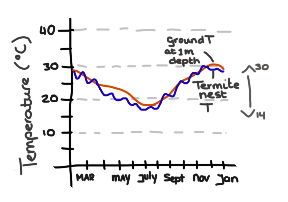
Nest temparature (blue) and ground temperature at 1m depth near nest (red) Drawing by M. Alleyne based on Tune r& Soar, 2008))

In addition, whereas induced flow might work well in tall buildings because the likelihood of a boundary layer gradient between locations is pretty high, it has been shown that induced flow rarely operates in termite mounds, even open-chimney mounds, since they are commonly only about 6 ft tall. There is also no evidence that mound ventilation and nest ventilation are indeed linked. How respiratory gases are moved from the nest to the mound, and fresh air from the outside, through the mound, to the nest, is not well understood.

Now the Eastgate Centre, and other large buildings since then, accomplished what they set out to do – they saved in construction costs (HVAC systems are very, very expensive) and they save on operating costs, all while keeping the inhabitants comfortable. It probably does not matter that they were based on incorrect science, but it does matter that the misconception gets repeated over and over again. If biomimicry and/or bioinspiration want to be considered legitimate fields of study, and not just a feel-good endeavor, then the science that the field is based on has to be solid.

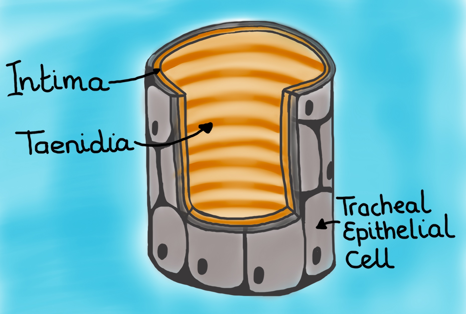
The termite mound should still inspire developers and architects because at it turns out, if we view the termite mound as the analogue of our own, respiratory system (lungs) then we still should be able to design “breathing” buildings that have that walls serve more as membranes rather than barriers.

Turner and Soar not only took relatively simple measurements of the temperatures, they also made plaster casts of the tunnel network of M. michaelseni mounds. They then created horizontal slices of the plaster filled mounds for easier recreation of future 3D models.

Based on these models, which showed actually very little continuous mixing between the air coming down from the above-ground structure and the air from the underground nest, they propose that the termite mound of M. michaelseni is in fact a functional analogue of a lung. And much like the lung the termite mound is far more complex than the simple models we have been using (for an excellent explanation comparing functional organization of both lungs and termite mounds please refer to Turner and Soar, 2008).

Comparison of the functional organization of mammalian lungs and the termite mound. There are areas of forced convection (large tidal flows) in red, areas where smaller tidal flows dominate creating mixing diffusion-convection, and then the areas where diffusion dominates.

Comparison of the functional organization of mammalian lungs and the termite mound. There are areas of forced convection (large tidal flows) (in red), areas where smaller tidal flows dominate creating a mix of diffusion-convection (in blue), and then the areas where diffusion dominates (in green). (Drawing by Marianne Alleyne, based on Turner & Soar, 2008)

This complexity opens up new avenues for bioinspired design based on the termite mound. Termites erect walls that are actually interface directly with the outside and indoor environments – the walls themselves let gases and energy through, it does not form an impenetrable barrier. Buildings that are designed based on correct termite mound architecture should incorporate porous walls and cladding (~skin) systems that incorporate tubes/tunnels through which air and energy can flow. Maybe we can design living buildings that are part of our extended physiology, as much as the termite mound is part of the living-system called the termite colony.

Resources:

I cannot recommend the Turner and Soar paper from 2008 enough. It is a wonderful read. The authors touch on more topics than I mention here (homeostasis, for instance). And again, their debunking of previously held beliefs is done in a way that should be emulated.

  • J. Scott Turner and Rupert C. Soar. 2008. Beyond biomimicry. What termites can tell us about realizing the living building.
  • The same authors also wrote a book chapter on a similar topic. But I have not yet been able to locate: Beyond biomimicry. What termites can tell us about realizing the living building. Chapter 15 in: Industrialised, Integrated, Intelligent Sustainable Construction. ISBN 978-0-86022-698-7. Ian Wallis, Lesya Bilan, Mike Smith & Abdul Samad Kazi (eds). I3CON/BSRIA. London. pp 233-248.

J. Scott Turner has also converted this work into very informative video lectures, such as:

  • Learning from nature: Termite mound lungs and the implications for breathing mines.

For great footage of how the termite mound models were created check out these videos.

I am not, by a long shot, the first to point out how the termite mound is NOT (yet) the perfect poster-child for bioinspiration and biomimicry. Here are two other blog-posts that discuss this very topic:

Additional pictures of the Eastgate Centre building in Harare Zimbabwe, click here.

Note: I would like to thank Adrian Smith who a few years ago made me aware of the work by Turner and Soar.

Going Micro-Tubular

Going Micro-Tubular

The ability to manufacture devices at the very small, or micro, scale brings with it interesting applications possibilities and surprising challenges.  For instance, certain physical phenomena may not be generally encountered with doing macro scale experiments on the lab bench but are dominant when experiments are done with a micro scale “lab-on-a-chip.” The field of microfluidics deals with the behavior and manipulation of fluids, both liquids and gases, that are constraint to a small scale (1 to several hundred micrometer). The physical properties of fluids at this scale are particularly interesting and could possibly be exploited.  For instance, fluids at a certain scale are no longer practically compressible.  Also, it is difficult to maintain pressure or pressure-driven flow within a micro scale fluidic system. One example is the phenomenon of back-flow whereby fluid moves in one direction when pressure is applied only to have it reverse and flow back when the pressure is released.  Due to the physics of scaling laws, these various non-intuitive phenomena are less severe when transporting gasses than liquids in a micro scale device.  For instance, gravity has no influence on gasses and diffusion of gasses at the micro scale is VERY fast.  Therefore, gas species can mix through diffusion with simple laminar flows at low Reynolds numbers.

In nature microscale liquid and gas flow is quite common. Maybe engineers should look at insects to see how fluids can most effectively be moved around at the microscale. The insect’s respiratory system, for instance, uses microscale flow transport to achieve an absolutely essential physiological function.  The respiratory system at first glance seems so simple – just some gas-filled tubes. At the same time it seems so foreign – no breathing through mouth or nose, no lungs or blood or hemoglobin involved (at least not at first glance). The respiratory system is also elegant in its simplicity through ingenious feedback loops – fine-tuning through direct nervous innervation or by sensing chemical compositions of the blood (called hemolymph in insects).

This system that is key to maintaining homeostasis is also as varied as insects are varied. Ingenious adaptations are seen in insects that live in different habitats – within soil, in water, in arid climates, in cold climates, within a host, while pupating, while flying, while walking, etc.

In this post I’ll focus mostly on the “generalized” terrestrial insect.

The insect’s tracheal system

The insect’s respiratory system’s main functions are to carry oxygen and carbon dioxide to and from the tissues. The gas-filled tubes, called tracheae, start at the spiracles, which are paired openings in the cuticle that can occur in almost every or only a few segments (depending on the insect species). The spiracles are innervated by the nervous system and can be open and closed through muscular action.

Spiracles

Spiracles of a lepidopteran larva. Two are visible in this picture; they appear as black openings surrounded by yellow ring. (Picture by Geoff Gallice)

Inside the insect the tracheal systems starts out as two or more longitudinal trunks, which further into the insect branch into smaller and smaller channels. Tracheae range in diameter from a few mm to 1 μm. The smallest branches are called tracheoles, which range from 1 to 0.1 μm in diameter. The tracheoles’ tips are in contact with cells and are often fluid-filled. This means that at the tips of the tracheoles oxygen has to cross first a liquid, then the tracheolar walls, then cross the plasma membrane of the cell and finally move through the cytoplasm of the cells to reach the mitochondria. Carbon dioxide makes the same trek, but in the opposite direction.

Tracheal System

Tracheal System (Drawing by Marianne Alleyne)

TrachealSystem

Cross-section of the tracheal system of “general” insect. Other insects may have more than 2 longitudinal trunks and/or air sacs and/or no spiracle functional in a particular segment. (Drawing by Marianne Alleyne)

Cross section of tracheal tube. A tracheal tube is comprised of an epidermal outer layer which bathes in hymolymph, an inner intima layer, and taenidia which reinforce the intima layer much like a car's radiator hose is reinforced by coil of steel. (Drawing by Marianne Alleyne)

Cross-section of tracheal tube. Shown is an epidermal outer layer bathing in hemolymph, an inner intima layer, and taenidia, which reinforce the intima layer (much like a car’s radiator hose is reinforced by coil of steel). (Drawing by Marianne Alleyne)

In order to get oxygen to move into the cells and carbon dioxide to move out of the body, all while limiting energy use, insects rely on diffusion and convection.

  • Diffusion: passive movement of molecules down their concentration gradient.

Oxygen diffusion rate is more rapid in air than in liquid, and because the tracheal system consists mostly of air-filled tubes the gas-exchange by diffusion is extremely rapid and quite substantial.  While the cells use the oxygen, the partial pressure of oxygen decreases near the tips of the tracheoles, which then draws in more oxygen from the tracheae and the air outside of the insect. Diffusion is of critical importance at the tips of the tracheoles, but it can only work at micrometer distances since at this point the oxygen has to move through a liquid. This is why almost every cell in the insect is at close proximity to a tracheole tip. The cells that require high respiratory rates (flight muscle cells, for instance) may even have multiple tracheole tips invaginating the cell wall.

The prominent Danish physiologist August Krogh devised an early model (1920) of grasshopper respiration that was based on oxygen partial pressures and suggested that simple diffusion could provide adequate gas exchange in insects. This claim had a huge impact on the field early and quickly became dogma, despite some contemporary contrary evidence. In 1964 “a next-generation Dane” Torkel Weis-Fogh showed that in larger or more active insects the distance between the spiracles and the tips of the tracheoles was too great and that diffusion was insufficient if the insect was active. So what other mechanisms do insects use to move oxygen and carbon dioxide through the tracheae?

  • Convection: bulk movement of a gas or a liquid driven by differential pressures.

Compared to diffusion, convection can achieve greater gas exchange rates over longer distances. The trade-off is that it requires more work and thus more energy.

    • One type of convective air-movement seen in insects is, confusingly, termed suction ventilation. This movement of air is primarily achieved by opening and closing, or shielding differentially, a subset or all of spiracles at different parts of the body. This creates a pressure difference between the spiracles and the tracheal ends and thus creates passive airflow.
    • Autoventilation is achieved by using wings or legs to pump air through the body.
    • Abdominal pumping, using the muscles in the abdomen to pump air.
    • Another, more important, type of convective air movement is achieved by the tracheae (tracheal compression), and their associated air sacs (tracheal sections that are not reinforced by taenidia), collapsing and inflating.
Silkworm Bombyx Mori Spiracle and Trachea 100X mag

Digital image of silkworm Bombyx mori trachea with taenidea (100X mag) by Paul Joseph, Birmingham, UK.

With novel imaging and measurement techniques researchers have shown that rhythmic tracheal compression is quite common.  Synchrotron x-ray imaging has been used since 2003 to visualize tracheal structures in living insects. We can now see that muscles compress the exoskeleton of the insect, which results in air moving through the tracheae since the tracheae and air sacs are collapsing.  These studies, which are now linked with real-time flow-through respirometry, show that rapid cycles of collapse and re-inflation in a variety of insects’ tracheal systems – especially in the head and thorax regions – results in large changes in tracheal volume and are major components of ventilation in insects. Even the different ways the tracheal system can compress and re-inflate is probably as varied as insects themselves.

Must watch: Beetle Tracheae Collapsing (video 1, video 2, video 3). Supporting materials from Westneat, et al. (2003) Science V299 (5606), DOI: 10.1126/science.1078008

Using the insect’s tracheal system as inspiration for microscale flow devices

The impetus for this blog post was a recent paper I came across entitled: Selective pumping in a network: insect-style microscale flow transport, by Yasser Aboelkassem and Anne E. Staples.  The topic of microscale flow transport is of interest to engineers because there are many applications that need precise flow control of just microliters of gas or fluid. Microscale flow devices or microfluidic devices are already around us and are assumed to change research tools in the life sciences, aid drug discovery and chemical analyses, and revolutionize health diagnostics. Modern DNA sequencing systems (Fluidigm, Illumina, Roche (454), etc.) rely on microfluidics. “Droplet” analyses also rely on microfluidic technologies. Cheap paper-based microfluidic immunoassay tests will become important for healthcare in the developing world (Diagnostics for All). And those are just a few of the applications.

Insect tracheal systems have been of particular interest to engineers now that we are able to visualize this physiological process (moving fluid and air through tubes) in action.  Insects are able to move fluid and air with precision with minimal energy requirements and little input from outside of the body. All characteristics that would work well within a “lab-on-a-chip”.

Insects use contractions at very specific places along the tracheal system, which probably affords them more precise control than if just relying on the more crude pressure difference approach. Through these small, but targeted, compressions the airflow to specific areas of the body can be achieved. This is apparently not an easy task to achieve in small-scale devices, and one can appreciate that by imagining larger tubes.

even tho a mushroom didn't ask me to

Imagine a garden hose…(Image by zen Sutherland)

Imagine a garden hose. By stepping onto the hose at one point the fluid or air inside the hose will be displaced to either side. As soon as you step off the hose the fluid or air will rush back (=back-flow). How can one create directional flow of a particular speed or pressure? By stepping at multiple spots along the hose, sometimes closing the hose off completely and other times only partially. Stepping off of the hose according to a certain sequence will also result in a directional flow. Of course, this is just one hose. Now imagine doing this with hoses that are branching into thinner hoses, that have multiple openings, with many more feet that can step on and off of them…and then think of a miniaturized version of that and you will see the insect’s tracheal system.

This is what Aboelkassem and Staples tried to model in their paper. The fluid dynamics research group headed by Staples at Virginia Tech’s Department of Engineering Science and Mechanics is apparently closely aligned with the biomechanics research lab of Jake Socha at the same departments. The Socha lab is one of the labs that has used synchrotron x-ray imaging to study insect respiration. The theoretical and computational model proposed by Aboelkassem and Staples incorporates rhythmic wall contractions along a network of tubes (in this case an 8-armed network) without the need for valves. The model’s tubes/channels are squeezed at different places along the tube. The contractions were actuated to move with different time lags from each other. In this way the researchers showed that fluids can be transported and flow velocity, pressure and direction can be controlled.

Simplified insect physiological network modeled by Aboelkassem & Staples. The "trachea" can be collapsed at multiple points within the network (red arrows). The collapse motion protocol can be varied by including time phase lags. The result is precise movement of the liquid or gas.

Simplified insect tracheal network modeled by Aboelkassem & Staples. The “tracheae” can be collapsed at multiple points within the network (red arrows). The collapse motion protocol can be varied by including time phase lags. The result is precise movement of the liquid or gas (green arrows).

We have learned from insects that localized rhythmic tracheal contraction is an effective way to move gas through micro-tubes. Aboelkassem and Staples claim to have been inspired by the insect’s tracheal system to model “selective pumping in a network”. They show, by using theoretical (low Reynolds number) flow analyses and computation fluid dynamics, that the network might also enable fluids to be transported precisely into particular tubes without the use of mechanical valves – at least through an 8-armed tracheal system. It will be interesting to see if this research group will continue to follow the insects’ lead by adding more branches/channels, by adding air sacs (which seem to be very important in insects), by making the model 3D, by adding actively opening and closing ports/spiracles to the outside, and by incorporating the diversity of insect respiratory systems into different models. If done correctly, and by using microfabrication techniques that can make flexible micro- and nano-scale tubes, it is likely insects can make a contribution to the rapidly growing technological field of microfluidics.

Resources and References:

For a more detailed description of the insect’s respiratory system:

For description of how synchrotron x-ray imaging is used to study insect respiration:

  • Westneat, M. W., O. Betz, R. W. Blob, K. Fezzaa, W. J. Cooper and W. -K. Lee. (2003) Tracheal respiration in insects visualized with synchrotron x-ray imaging. Science V299, 5606, pp. 558-560. DOI: 10.1126/science.1078008
  • Westneat, M.W., J.J. Socha and W.-K. Lee. (2008) Advances in biological structure, function and physiology using synchrotron x-ray imaging. Annual Review of Physiology V70: 119-142. DOI: 10.1146/annurev.physiol.70.113006.100434
  • Socha, J.J., W.-K. Lee, J.F. Harrison, J.S. Waters*, Fezzaa, K. and M.W. Westneat. (2008) Correlated patterns of tracheal compression and convective gas exchange in a carabid beetle. Journal of Experimental Biology V211: 3409-3420. DOI: 10.1242/​jeb.019877
  • Socha, J.J., T. Förster and K.J. Greenlee. (2010) Issues of convection in insect respiration: Insights from synchrotron x-ray imaging and beyond. Respiratory Physiology and Neurobiology V173S S65–S73. DOI: 10.1016/j.resp.2010.03.013
  • Waters J.S.,W.-K. Lee, M.W. Westneat and J. J. Socha. (2013) Dynamics of tracheal compression in the horned passalus beetle. American Journal of Physiology:Regulatory, Integrative and Comparative Physiology V304(8) R621-7. DOI: 10.1152/ajpregu.00500.2012

Bioinspired microscale flow structures:

Special thanks to Dr. Paul Kenis for taking the time to discuss microscale flow transport with me. I appreciate his patience with me – clearly a novice. Bedankt!